Modeling the development of topographic maps in the brain
Primate vision research has shown that in the retinotopic map of the primary visual cortex, eccentricity and meridional angle are mapped onto two orthogonal axes: whereas the eccentricity is mapped onto the nasotemporal axis, the meridional angle is mapped onto the dorsoventral axis Theoretically such a map has been approximated by a complex log map. We attempt to model the development of the global retinotopic and orientation maps using a similar self organizing model.
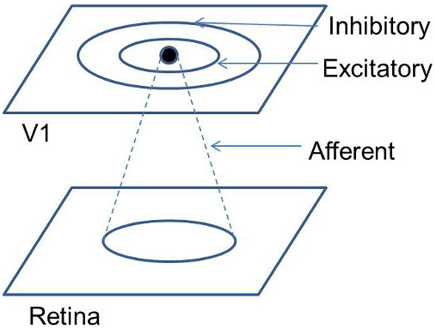
The architecture consists of a Laterally Interconnected Synergetically Self Organizing Map (LISSOM) with 2 layers; representing the retina, and the V1 respectively At each time step, each neuron in V1, combines the afferent activation along with its lateral excitations and inhibitions from the previous time step. The afferent, lateral excitatory and lateral inhibitory weights adapt based on a normalized Hebbian mechanism.
R. T. Philips and V. S. Chakravarthy, "The mapping of eccentricity and meridional angle onto orthogonal axes in the primary visual cortex: an activity-dependent developmental model," Frontiers in computational neuroscience, vol. 9, 2015 PDF.


