Research

RESEARCH

Research

A Study on the Activators and Targets of NF-κB in Human Cancer Cells

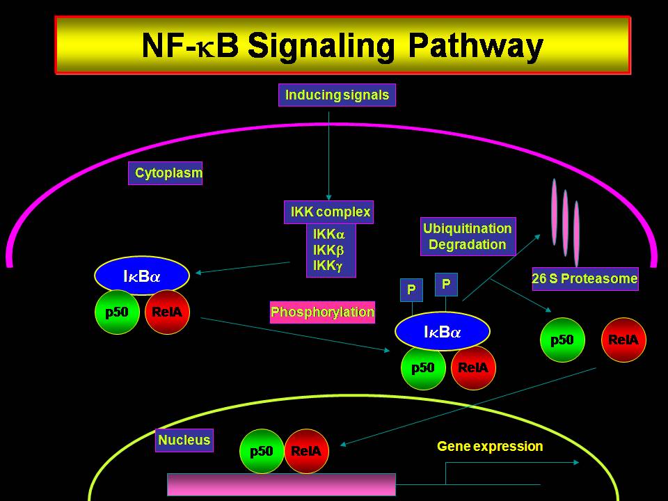
The ubiquitous transcription factor, NF-κB, is deregulated in many solid and hematological malignancies. Most of the tumor types and cell lines show constitutive activation of NF-κB that enables them to resist chemo and/or radio therapy by inducing the expression of anti apoptotic/survival factors. In addition NF-κB enhances the ability of tumors to metastasize as well as form new vasculature, which are critical for tumor survival. Since constitutive NF-κB activity correlates positively to the resistance of cancer cells to anti cancer agents, antagonizing NF-κB offers an immense therapeutic potential to improve the clinical response in these tumor types. Many studies have shown that downregulation of NF-κB sensitizes cells to therapy as well as inhibits the metastatic and angiogenic ability of different tumor types. Recently we have reported that NF-κB is constitutively activated in high grade squamous intraepithelial lesions and squamous cell carcinomas of the human uterine cervix. However, the mechanisms by which tumors maintain a sustained activation of NF-κB are not clearly defined. Since ex vivo tissue explants are not very much suited for mechanistic studies, the present proposal is aimed at identifying the factors responsible for the activation of NF-κB and their targets in human cervical cancers using in vitro model systems. Hence it is proposed to study the expression of activators of NF-κB and their targets which are often responsible for tumor initiation, progression, invasion and metastasis. It is proposed to test the hypothesis that cytokines and growth factors secreted by tumor cells as well as the surrounding stromal layer are responsible for constitutive activation of NF-κB. Organotypic raft culture will be used as a mechanistic model to test this hypothesis and it will also serve as an in vitro model for testing the efficacy of anti cancer drugs.

Cytomodulins in Oral Cancer Therapy

Cancer cells may lose their sensitivity and responsiveness to TGF-β. Oral cancer is the most prevalent type of cancer in India accounting for 40% of all cancers and it has a low survival rate. In cancerous tissues, apoptotic signalling by TGF-β is blocked by the loss of its anchoring receptor TβRII, as a sequel to dominant TNF-a activity induced by inflammation, radiation, viruses, and reactive molecules. NF-?B plays a major role in blocking apoptosis and does so partly by blocking TGF-β; signalling. Our hypothesis is that cytomodulins and CITAs (conformationally restricted analogues of cytomodulins), synthetic agonists of TGF-β signalling receptor TβRI will be effective as a novel therapy for oral and other cancers. Previous collaborative studies between Bhatnagar and Karunagaran have shown that cytomodulins and CITAs initiate TGF-β signaling in cells lacking TβRII. Cytomodulins inhibit the activation of TNF-a-induced NF-κB. CITAs inhibit both constitutive and induced activation of NF-?B. These compounds are not cytotoxic. In a preliminary clinical study topically applied cytomodulin was found to be rapid-acting and highly effective against squamous cell carcinomas, oral cancer lesions and in cancer therapy induced oral mucositis. Cytomodulins and CITAs are excellent therapeutic candidates for oral cancer. These compounds were invented by Co-PI, Rajendra Bhatnagar. Our specific aims in this collaborative study are (i) to determine the best cytomodulin/CITA candidates based on structure activity relationships (Bhatnagar), by (ii) examining the mechanisms of action of diverse peptides using in vitro models (Karunagaran), (iii) examine the therapeutic efficacy of the short-listed compound(s) in animal models (S.Nagini). Based on studies in the first three aims, (iv) we will conduct pharmacological and toxicological studies of the best candidate compounds at NIPER (being negotiated) (vi) expeditiously to obtain regulatory approval and conduct human testing. A coordinated multi-disciplinary effort will permit us rapidly to develop a safe and simple chemotherapeutic modality for treatment of oral cancer. The results from these studies will provide a basis for treatment of other NF-κB -involving diseases including various cancers, inflammatory diseases, skin diseases e.g. psoriasis, and Alzheimer syndrome.

A Functional Characterization of miRNAs in Human Cancer Cells

Cancer will be one of the major diseases in India by 2025. Initiation, promotion, malignant conversion, progression and. metastasis are the five major steps in cancer development. Combined interaction of both tumor suppressors and inducers is the cause for the formation of cancer. Even though several genes had been identified in human, the mechanism of cancer formation is yet to be ascertained. A recently identified class of non-protein coding small RNAs, micro RNAs (miRNA), may provide new insight in cancer research. miRNAs are short, non-coding RNAs that regulate gene expression by controlling the efficiency of mRNA translation. Many miRNAs exhibit altered expression levels in cancer, and scientific community is only starting to understand the functional consequences of the loss or gain of particular miRNAs to cancerous phenotype. miRNAs are also naturally occurring triggers of the RNA-interference pathway. miRNAs are expected to regulate more than 30% of all mRNAs post-transcriptionally. Several groups have studied the miRNA expression in cancer patients and found that miRNAs are differentially expressed in normal and cancer tissues. Some miRNAs may function as oncogenes or tumor suppressor genes. Examples of their roles in development and disease are rapidly accumulating but the biological functions of most of them are completely unknown. Here we propose to elucidate the functions of reported miRNAs in cancer cells in the following manner. 1. Over-expression of miRNAs 2. Knocking down miRNAs using genetic approaches 3. Silencing the miRNAs using antagomirs (single strand RNA) and siRNAs 4. Replacement of miRNAs (in case of tumor suppressor miRNAs). The outcome of these results will be compared with computational predictions of the target. Up- and/or down-regulated expression of the candidate miRNAs are a good approach to study the function of miRNAs in cancer pathogenesis. It will also be important to elucidate biochemical pathways that affect miRNA function as well as environmental and genetic factors that affect miRNA expression. Once this information is available, a rational approach can be taken to design new therapeutic strategies that aim to treat cancer.

Studies on TGF-β-mediated changes in gene expression and signaling pathways in human cervical cancer cells

Transforming growth factor-β (TGF-β) family includes a large number of structurally related polypeptide growth factors-TGF-βs, activins and bone morphogenetic proteins-that are important in homeostasis and repair of virtually all tissues. TGF-βs were identified as regulators of mesenchymal growth and differentiation, cell cycle arrest in epithelial and hematopoetic cells, extracellular matrix production and immunosuppression. We assessed the responsiveness of six human cervical cancer cell lines to TGF-β with p3TP-lux reporter assay and found that HeLa and SiHa cells were highly responsive to TGF-β. However, when pSBE4-BV/Luc reporter having 4 smad binding elements was used, only the SiHa but not HeLa cells showed Smad activation. Smad DNA binding activity was relatively more in SiHa than HeLa cells upon TGF-β treatment and the active complex contained Smad 2 and Smad 4. In MTT assays, HeLa cells treated with 5ng/ml of TGF-β for 24h showed proliferation whereas SiHa cells showed growth inhibition under the same conditions. TGF-β treatment resulted in G0/G1 arrest with reduction in S-phase only in SiHa cells and a chemical inhibitor of Smad activation (SB203580) blocked the growth inhibitory effect of TGF-β in SiHa while the proliferative response in HeLa was unaffected. TGF-β-induced translocation of phospho-Smad 2 was relatively less in HeLa than SiHa cells. MAPK activation occurred within 5 min and persisted up to 15 min upon TGF-β treatment in HeLa but was negligible in SiHa cells. TGF-β activated JNK in HeLa but SiHa cells showed a down-regulation of its activity. When an inhibitor of MAPK (U0126) was used, the TGF-β-mediated proliferative response in HeLa cells was completely abolished. SB203580 did not affect MAPK activation induced by TGF-β in HeLa cells. We reported for the first time an activation of TGF-β -dependent MAPK within 5 min of treatment that resulted in cell cycle progression in a cervical adenocarcinoma cell line, HeLa, which is presumably independent of Smad activation. Hence, in the present proposal it is planned to study smad independent mechanisms by down regulation of smads by SiRNA or antisense approaches. Studies on TGF-β-mediated changes in MAPK, Rho, Rac and Rock signaling pathways in human cervical cancer cells will also be undertaken. Micro array approaches to study differential regulation in cervical cancer cells are also planned.

Search The Literature
Search Pubmed


Contact Email
Email : karuna@iitm.ac.in

Contact Phone
Call : +044-2257 5118

Social Media
Skype : karuna1955
Facetime id : karuna@iitm.ac.in
Twitter id : karunagarand
linked in id : karunagaran-devarajan-4835288
Powered by Dreamstech.co